 White dot syndromes | Eye News
What is this white spot on my eyeball? There are only a few causes of white spots in the eye, and most are easily treatable. The most common reasons are the corneal ulcers and the pinguecula. White points in the eyeball can vary in severity. Some may be unperceptable while others may cause a lot of discomfort. Eye problems of any kind can cause long-term damage to vision. Even if discomfort is minimal, always seek medical advice if a white point appears in the eye. In this article, we observe the diagnosis and treatment of white spots in the eye. We also analyze how to prevent them, and likely results. Conditions that can cause a white point to form in one eye include: corneal ulcersThe corneal ulcers can lead to . They can also lead to blindness if not treated. The ulcers occur when the cornea is damaged. The causes of damage may include: If something breaks through the surface of the cornea, an infection may develop. Germs that may cause infection in the cornea include: Other conditions that may cause corneal ulcers arePingueculas Pingueculas is another common cause of white spots in the eye. They can occur when the eyes: Pinguecula stains are white or yellow and consist of fat or protein deposits. They appear in the conjunction, which is the transparent cover of the white part of the eyeball. These stitches are usually irregular in shape and usually form by the eye closest to the nose. Cancer Cancers can also form in the eyeball. These include: Ocular cancers, as you know, are . Some cancers have environmental causes, such as sun exposure. In other cases, they may occur due to a person's genetics. A corneal ulcer, pinguecula, or eye cancer may share some common symptoms, such as:Each cause also has some unique symptoms. Body Ulcer Symptoms Pinguecula Symptoms Although the pingüecula may occur without additional symptoms, they may be accompanied by: Symptoms of eye cancer Eye cancers may initially appear to be minor conditions. An eye test should collect any signs of eye cancer, including: Anyone who has an eye problem that is not clarified in one day or two should look for treatment. It is essential to see a doctor if there is: A doctor may refer to someone to an ophthalmologist or unptometrist. These are eye specialists who can perform a full range of tests. An eye doctor will examine the eye and ask about recent injuries. They can also perform a test of the cutting lamp. To do this test, the ophthalmologist or optometrist drops a dye called fluorescein in the eye, which they will examine using a special microscope. If your eye doctor suspects an infection, you may take a small amount of tissue for the test. This is called biopsy or culture. Doctors can also identify pinguecula by examining the eye or using fluorescein. This will usually be enough, but a doctor may request a biopsy if they are not sure. A doctor may diagnose eye cancer with the following tests: Treatment of corneal ulcers A doctor will remove any foreign body from the eye and then treat the damage. A person should not use contact lenses during treatment and recovery, even if they are not the cause of the corneal ulcer. The drops are one of the most common treatments for corneal ulcers. The best type of eye drop to use depends on the causes. The range of eye drops includes: Pain relief medicine is available in the form of eye drops and pills. Once an infection has been clarified, people can use eye drops of steroids to treat any scar that has been formed. However, steroids can make things worse if used before an infection is clarified. Substitutes of tears can help if the eyes do not produce enough moisture. If the damage to the cornea is severe, doctors can replace part or whole cornea with a new donor cornea. Treatment of the brushes Eye drops and ointments can usually treat the pinguecula. If a pinguecula affects a person's vision or causes severe discomfort, it may require surgery to remove them. Laser treatment is becoming more common. Ocular Cancers Treatments for eye cancers depend on the type and stage of the . Treatment methods include: The best way to prevent white spots from developing in the eyes involves taking care of the eyes. The following steps can help protect the health of your eyes: If someone has one, they should avoid touching their eyes. People who wear contact lenses should always follow the guidelines for use. It is essential to clean the lenses with the right solution, remove them regularly, and replace them if they are damaged or poorly adapted. The perspective of the white points in the eye can vary a lot. A white place is most likely to be a short-term condition. In terms of treatment, results after diagnosis are excellent. Although the chances of a white place being cancer are low, potential risks are high. White points in the eye tend to be easily treatable. An early diagnosis will help limit the impact. It is therefore essential to talk to a doctor if there are signs of white spots in the eye. Good hygiene practices are always recommended and will help prevent many eye problems. Be careful to protect your eyes against the sun, particles in the air and bacteria. Last medical review on October 12, 2018Most recent newsRelated coverage
IMAGES
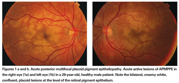
Recognizing the 'White Dot' Syndromes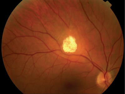
White Spot' Draws Attention for 30 Years
AI trained to spot heart disease risks using retina scan | Ars Technica
The white dot syndromes
White dot syndromes | Eye News
MEWDS (Multiple Evanescent White Dot Syndrome) - Retina Associates
The white dot syndromes
Pin by Mustafa Aksoy on Coats Disease.... Arm yourself with knowledge! | Eye health, Vision eye, Retina surgery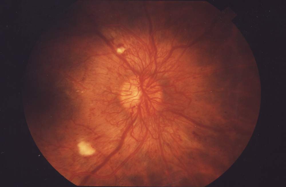
Cotton wool spots - Wikipedia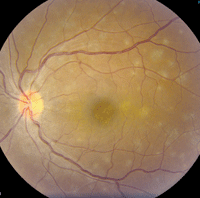
The Eye is Dotted
Characterizing the White Dot Syndromes - Retina Today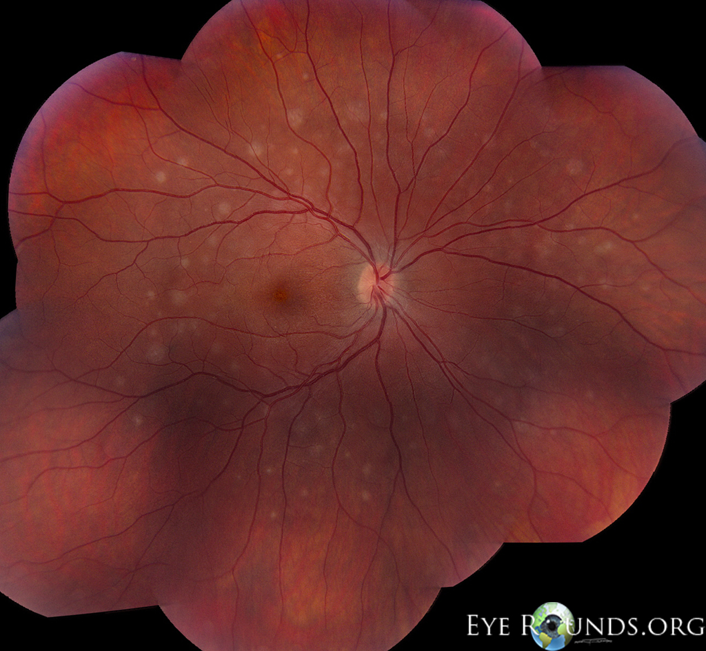
Multiple evanescent white dot syndrome (MEWDS)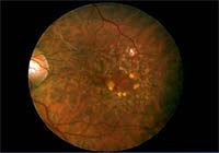
Conditions: Macular Degeneration | Eugene Eye Care
Characterizing the White Dot Syndromes - Retina Today
White-Centered Retinal Hemorrhages | Consultant360
Roth Spots: Pictures, Causes, Diagnosis, and Treatment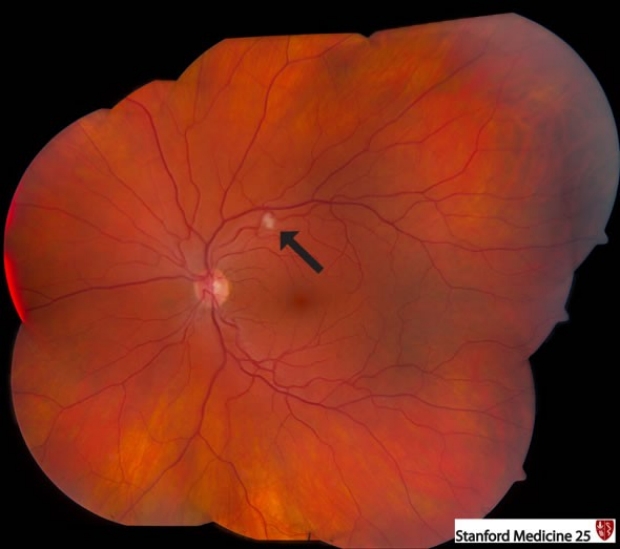
Fundoscopic Exam (Ophthalmoscopy) | Stanford Medicine 25 | Stanford Medicine
Areas of Fundus Whitening: White or Yellow Spots | Ento Key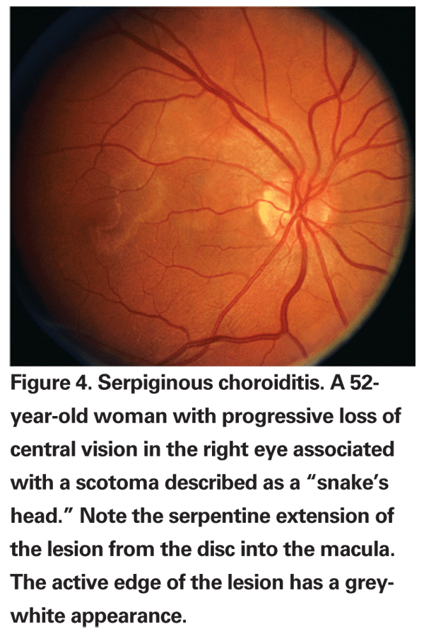
Recognizing the 'White Dot' Syndromes
Retina Eye Specialists - www.retinaeye.com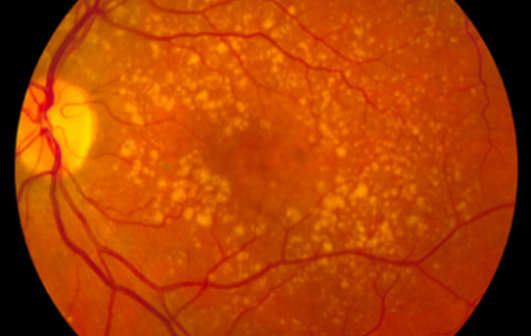
AI trained to spot heart disease risks using retina scan | Ars Technica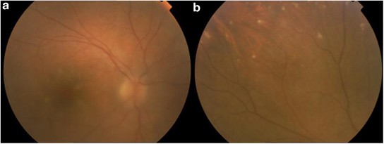
White dots in the eye fundus revealing Hodgkin's lymphoma | Eye
Retinal Signs - Angeles Vision Clinic
Nevus (Eye Freckle) - American Academy of Ophthalmology
Diabetic Retinopathy - Arleo Eye Associates - Arleo Eye Associates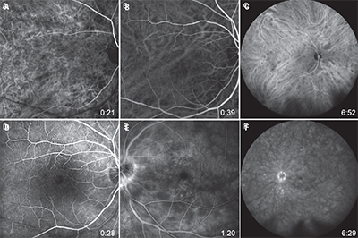
Which White Dot Syndrome Is It?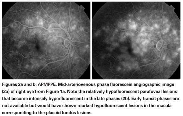
Recognizing the 'White Dot' Syndromes
FULL TEXT - Bilateral exudative multifocal retinal detachment: An unusual presentation of accelerated hypertension with obstructive uropathy - International Journal of Case Reports and Images (IJCRI)
Cotton Wool Spots : Ophthalmoscopic Abnormalities : The Eyes Have It
Macular Edema - The American Society of Retina Specialists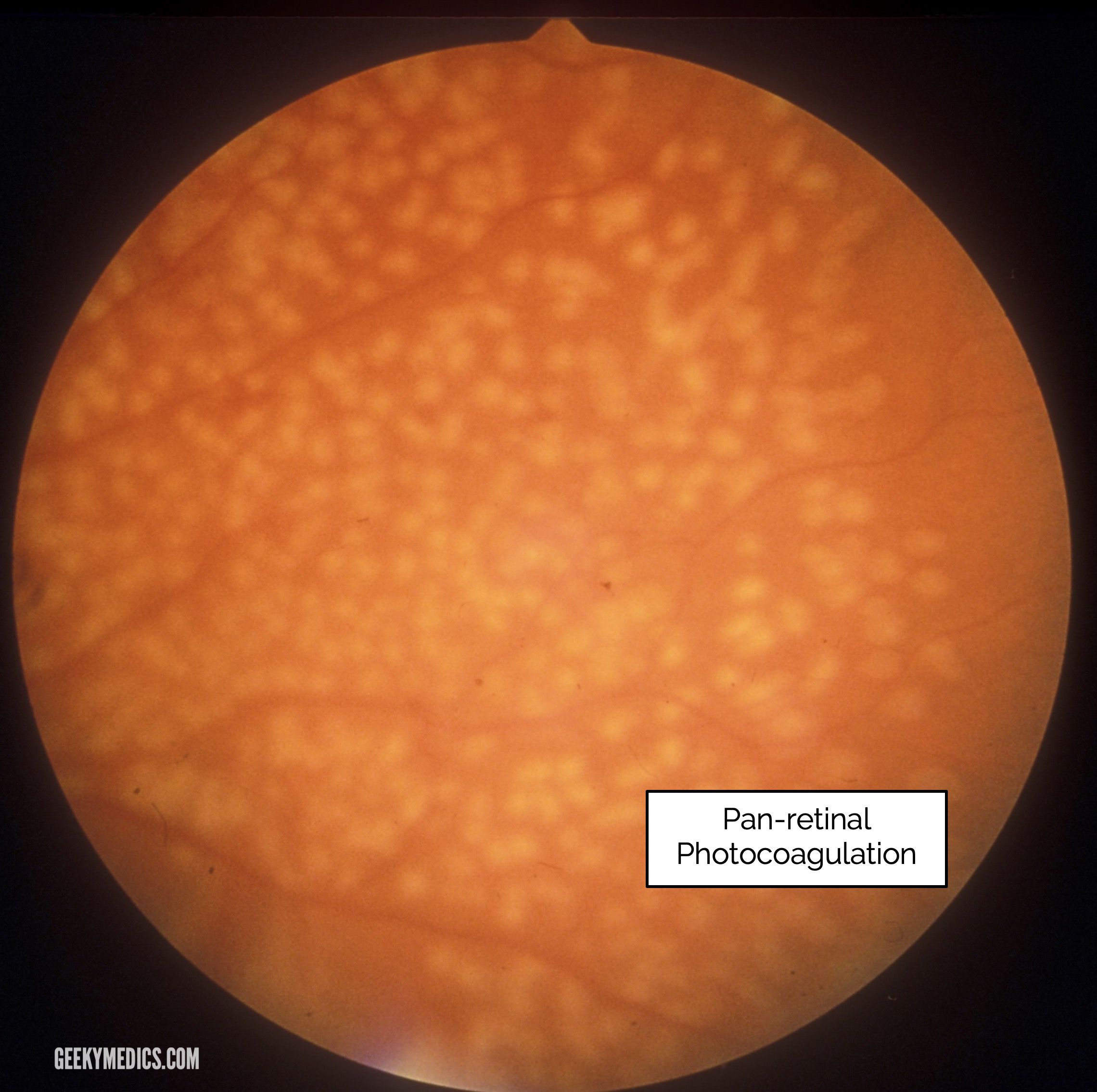
Fundoscopic Appearances of Retinal Pathologies | Geeky Medics
White dot syndromes | Eye News![Full text] Multiple evanescent white dot syndrome associated with retinal vasculi | IMCRJ Full text] Multiple evanescent white dot syndrome associated with retinal vasculi | IMCRJ](https://www.dovepress.com/cr_data/article_fulltext/s88000/88639/img/fig2.jpg)
Full text] Multiple evanescent white dot syndrome associated with retinal vasculi | IMCRJ
ISOPTIK : Retina
A sample retinal image with cotton wool spots and hemorrhages. | Download Scientific Diagram
Retinal Physician - Management of Cotton-wool Spots in Retina
Why cotton wool spots should not be regarded as retinal nerve fibre layer infarcts | British Journal of Ophthalmology
White dot syndromes - Neuro-Ophthalmology
Diabetic Retinopathy | Scott E. Pautler, M.D. Tampa
Areas of Fundus Whitening: White or Yellow Spots | Ento Key
 White dot syndromes | Eye News


![Full text] Multiple evanescent white dot syndrome associated with retinal vasculi | IMCRJ Full text] Multiple evanescent white dot syndrome associated with retinal vasculi | IMCRJ](https://www.dovepress.com/cr_data/article_fulltext/s88000/88639/img/fig2.jpg)


Posting Komentar untuk "white spots on retina"